About

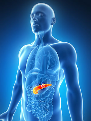
Pancreatic cancer begins when abnormal cells in the pancreas grow and divide out of control and form a tumor.
The pancreas is a gland located deep in the abdomen, between the stomach and the spine. It makes enzymes that help digestion and hormones that control blood-sugar levels.
Organs, like the pancreas, are made up of cells. Normally, cells divide to form new cells as the body needs them. When cells get old, they die, and new cells take their place.
Sometimes this process breaks. New cells form when the body does not need them, or old cells do not die. The extra cells may form a mass of tissue called a tumor.
Some tumors are benign. This means they are abnormal but cannot invade other parts of the body.
A malignant tumor is called cancer. The cells grow out of control and can spread to other tissues and organs.
Even when the cancer spreads to other areas of the body, it is still called pancreatic cancer if that is where it started. Pancreatic cancer often spreads to the liver, abdominal wall, lungs, bones and/or lymph nodes
(Pancreatic cancer-Facing pancreatic cancer
Current treatment methods
Pancreatic cancer treatment depends on the stage of the disease and the patient’s general health. Patients may get standard (approved) treatments or take part in clinical trials.
Standard treatments are surgery, chemotherapy and radiation. Clinical trials study new treatments. The Pancreatic Cancer Action Network strongly recommends clinical trials at diagnosis and during every treatment decision.
What type of pancreatic cancer patients is particularly well suited for HIFU?
Patients with locally advanced pancreatic cancer and with unresectable pancreatic cancer. In patients with advanced pancreatic cancer and otherwise limited treatment options, HIFU resulted in significant early and long-lasting pain relief and tumor size reduction over time independent of metastatic status. Clinical data suggest an additional potential survival benefit.
*Please be noted the above criteria is only for reference, whether a patient can be applied HIFU shall be further confirmed by HIFU doctors after diagnosis.
How does HIFU work for pancreatic cancer (the advantages)?
HIFU can be used for local ablation of advanced pancreatic cancer. As the temperature in the target region rises to over
80°C, the main effect of HIFU is a thermal ablation, which causes regulation necrosis and thus a volume reduction over time. In conclusion, noninvasive US-guided HIFU for advanced pancreatic cancer is a well-tolerated, low-risk procedure with beneficial clinical effects and significant reduction of pain burden independent of metastatic status as well as consecutive improvement of patient quality of life. As HIFU also results in a significant reduction in tumor volume, the procedure may be considered when tumor bulk reduction is desirable, e. g. in the context of other therapies. Survival data suggest a possible survival benefit. HIFU can be of value in this patient population with otherwise limited treatment options. To validate our findings, we are currently performing a randomized controlled clinical trial comparing standard palliative therapy alone with standard therapy plus HIFU in late-stage pancreatic cancer.
Clinical evidence
According to Clinical Effectiveness and Potential Survival Benefit of US-Guided High-Intensity Focused Ultrasound Therapy in Patients with Advanced-Stage Pancreatic Cancer, in 84% patients among 50 with late-stage pancreatic cancer undergoing HIFU in the study, significant early relief of cancer-induced abdominal pain was achieved by HIFU independent of metastatic status; it persisted during follow-up. Tumor volume reduction was 37.8
± 18.1% after 6 weeks and 57.9 ± 25.9% after 6 months. The median overall survival and progression-free survival were 16.2 and 16.9 months from diagnosis.
Safety
The most common adverse events(Class A) of HIFU treatment for gyenecological diseases are vaginal discharge, skin burn and etc. Serious adverse events (Class C&D) are less than 1%.
A multi-center study in China: 2011 to 2017, 27,053 patients with benign uterine diseases were treated with HIFU in 19 centers in China. Among them, 17,402 patients had uterine fibroids, 8434 had adenomyosis, 876 had caesarean scar pregnancies, and 341 had placenta accreta. After HIFU treatment, 13,170 adverse events were observed. Based on society of interventional radiology classification system, these adverse events were classified as Class A (47.5030%), Class B (0.7947%), Class C (0.3327%), and Class D (0.0518%). The rate of major adverse effects (Class C&D) was 0.3844%. Major adverse effects include skin burn, leg pain, vaginal discharge or bleeding, urinary retention, acute cystitis, intrauterine infection, bowel injury, acute renal failure, deep vein thrombosis, pubic symphysis injury, post-HIFU thrombocytopenia, sciatic nerve injury, and hydronephrosis. In 2011, the annual rate of major adverse effects was 0.9565%; the incidence decreased to 0.2852% in 2017. it concluded that “based on the results with low rate of major adverse effects from multiple centers, we concluded that HIFU is safe in treating patients with benign uterine diseases. With development of this technique and more experience on the part of the physicians, the rates of the major adverse effects will be further lowered”.
Adverse effect analysis of high-intensity focused ultrasound in the treatment of benign uterine diseases. Liu Y, Zhang WW, He M, Gong C, Xie B, Wen X, Li D, Zhang L.
Int J Hyperthermia. 2018;35(1):56-61. doi: 10.1080/02656736.2018.1473894. Epub 2018 May 24.
Cases for pancreatic cancer treatment

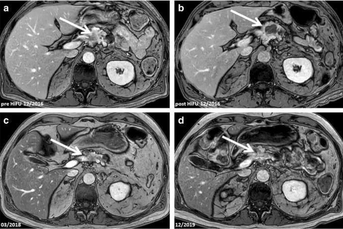
Fig.1 A 70-year-old male patient with non-resectable locally advanced adenocarcinoma of the pancreatic body and tail (UICC stage III) was treated by HIFU. No simultaneous chemotherapy was given to the patient because of intolerance.
Notes: a: Tumor (arrowheads) before HIFU treatment. b: One day after HIFU, treated tumor regions showed no contrast enhancement indictive of effective ablation. c, d: Sustained tumor reduction in MRI studies at the c 1.5- and d 3-year follow-up.

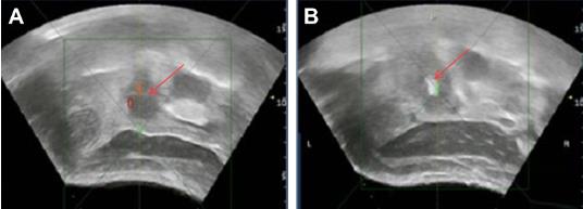
Fig.2 Grayscale changes in treated pancreatic cancer tissue on real-time US images during HIFU.
Notes: A: US image obtained during HIFU treatment showing a pancreatic cancer lesion present in the head of the pancreas (red row). B: US image obtained immediately after HIFU treatment showing obvious hyperechogenicity of the treated pancreatic tumor (red row).
Patient stories
After being diagnosed with inoperable pancreatic cancer, Mr. Du came to Chongqing Haifu Hospital seeking for help with his son. In Mr. Du’s follow-up consultation in May after the successful HIFU treatment three months ago, his son said, “it was a more than ten’s hour drive from Hebei to Chongqing with my 68-year-old father, because Chongqing Haifu Hospital is reliable!” Prof. Zhang Lian told them, “According to the MRI, the ablated lesion is basically absorbed. With pain relief and sound diet, sleep and mood, we can say that the HIFU treatment is effective and produces expected outcomes.”